FibroNest
The First Single-Fiber, High-Resolution
and Fully Translational Quantitative AI Digital Pathology image analysis Method for the Phenotypic Quantification of Fibrosis and it’s Associated Features
~ 9 Phase 2 Studies
~ 2 Phase 3 Studies
~ 15 Fibrotic Conditions
~ +22 Validated Rodent Models
~ 140 Pre-clinical Studies
~ 2 Organoid Models
~ 6.3 K Clinical Images
~ 11K Images
~ 78T Single Fibers Data
~ 9 Phase 2 Studies ~ 2 Phase 3 Studies ~ 15 Fibrotic Conditions ~ +22 Validated Rodent Models ~ 140 Pre-clinical Studies ~ 2 Organoid Models ~ 6.3 K Clinical Images ~ 11K Images ~ 78T Single Fibers Data
USINg FIBRONEST, PHARMANEST provideS HIGH RESOLUTION, SINGLE FIBER FIBROSIS PHENOTYPIC QUANTIFICATION services FROM DIGITAL IMAGES OF STAINED SLIDES FOR clinical and pre-clinical pharmaceutical DRUG development PROJECTS
FibroNest is a Research Only Use product for now and can be used for pre-clinical projects, for research clinical projects or as an exploratory outcome
Click here to request a Consultation
MULTIVENDOR COMPATIBILITY
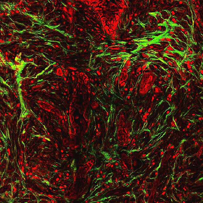
FibroNest is compatible with all kinds of Digital Images, ranging from FDA-Approved WSI scanners to Fluorescence and Second Harmonic Generation microscopes
SINGLE-FIBER
FROM ALL KINDS OF COLLAGEN STAINED IMAGES
Collagen-stained digital images (PSR, MTC, IHC) are quantified by FibroNest at the fiber level, with full spatial localization
TRANSLATIONAL
FibroNest’s engine is fully translational from discovery to clinical development, and is validated for multiple fibrosis conditions, including liver, lung, kidney, skin ….


